A forest inside your head
A three-year-old asks why the moon follows the car. A four-year-old remembers a song they haven't heard in weeks, balances on one foot, or tells a story about something that happened yesterday.
All of this happens because of a network of billions of cells inside their head. Cells that send messages, respond to one another, and change as a child learns.
Most of us know about neurons, the cells that carry information through the nervous system. A brain made entirely of neurons wouldn't work. There are other cells living among them, doing jobs that neurons cannot do alone.
What does it take to keep a brain working?
The trees
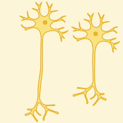
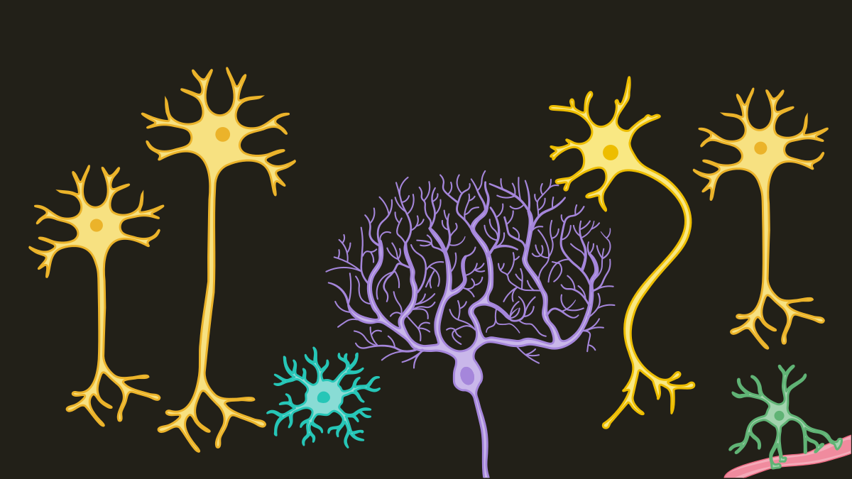
Look at a neuron under a microscope and you'll see something that resembles a tree. Branches spread out from its cell body, receiving messages from other cells, while a long extension called an axon carries electrical signals away.
Some neurons have elaborate branches. Purkinje cells, found in the cerebellum at the back of the brain, spread their branches into a flat fan that looks almost like a pressed leaf. Each one receives an enormous number of signals, helping the cerebellum coordinate movement and balance.
When a child learns to hop on one foot, these cells are among those helping the brain make the adjustments needed to stay upright.
Neurons communicate at connections called synapses. At most synapses, one neuron releases chemical messengers into a tiny gap, where the next cell detects them and responds.
A single neuron can form connections with thousands of others. Together, they create networks that allow information to move through the brain. These networks are always changing.
A forest needs more than trees
For a long time, scientists thought the other cells surrounding neurons were little more than structural support. They called them glia, from the Greek word for glue.
It turned out that the brain's supposed glue was doing some remarkable things.
Astrocytes, named for their star-like shape, reach toward neurons and blood vessels. They help supply neurons with energy, regulate the chemicals surrounding them, and maintain the blood-brain barrier, which helps control what enters brain tissue from the blood.
Oligodendrocytes wrap parts of neurons' axons in a fatty coating called myelin. Like insulation around an electrical wire, myelin helps electrical signals travel quickly and efficiently. This wrapping continues to develop throughout childhood and beyond.
Deep inside the brain, ependymal cells line chambers filled with cerebrospinal fluid, the clear liquid that cushions the brain and spinal cord. Tiny hairs on these cells beat back and forth, helping keep the fluid moving.
None of these cells carries messages in quite the way neurons do. But without them, the environment neurons depend on would be very different. There's another type of glial cell with a particularly surprising job.
The gardeners
Microglia are the brain's resident immune cells.
Under a microscope, they look like tiny branching creatures. Their thin branches are constantly moving, surveying the surrounding tissue for signs of injury, infection, or damaged cells. When something needs to be cleared away, microglia can engulf and digest it. Protecting the brain isn't their only job.
During early development, the brain produces an enormous number of connections between neurons. Not all of them will be maintained. As children grow, some connections become stronger, while others weaken or disappear.
Microglia help shape these developing networks by participating in the removal of certain synaptic connections, a process called synaptic pruning.
Think of a forest growing thick with branches. Growth isn't only about adding more. Sometimes branches are removed, allowing the remaining structure to change.
Something similar happens in the developing brain. Its networks are continually being reorganized, with neurons, microglia, and other cells all contributing to the process.
A child's brain isn't just getting bigger or collecting more information. The connections between its cells are changing too.
Where the forest begins
Long before a child learns to speak or walk, the brain is already taking shape. During pregnancy, neural stem cells divide and give rise to the cells that eventually become neurons and glia. Many newly formed neurons travel to specific locations, grow their branches, and begin making connections.
Most of the brain's neurons are produced before birth. The networks they form continue developing for years afterward.
Connections strengthen or weaken with experience, myelin continues forming around axons, and different parts of the brain mature at different rates.
The accompanying brain map shows some of these regions, from the cerebellum that helps coordinate movement to the areas involved in seeing, hearing, remembering, and feeling. Although different regions have specialized roles, they work together through networks of connected cells.
Still growing
The next time a child remembers a song, learns a new movement, or asks another question you weren't expecting, think about the forest inside their head.
Neurons are exchanging messages, oligodendrocytes are wrapping axons, astrocytes are maintaining the environment around them, and microglia are helping shape the connections between cells.
None of them could do the whole job alone.
The eight cards in this pack show some of the cells that make up that forest, each with a different shape and a different role. Together, they make possible the things a child does every day, often without any of us thinking about what's happening inside. The forest keeps changing, long after childhood ends.
Pack 10, Inside Your Brain, was made with Christy Kestner, PhD, neuroscientist, neuroimmunologist, and founder of Brain & Beyond.